
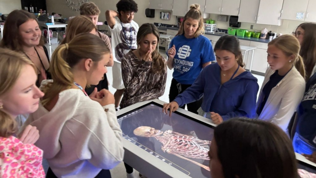
Medical colleges can integrate virtual dissection by mapping it to anatomy learning outcomes, using it before and after cadaveric sessions, and linking it to assessment and clinical cases. The goal is not to replace established anatomy teaching, but to give students more opportunities to explore, revisit, and apply anatomical knowledge.
In clinical practice, anatomy is understood through relationships rather than isolated structures. A surgeon needs to know where a nerve, vessel, muscle, or organ sits—but also what surrounds it, what lies beneath it, how it relates to neighbouring structures, and how those relationships can vary.
Cadaveric dissection remains fundamental to this kind of understanding. It lets learners work through tissue planes, depth, spatial relationships, and anatomical variation. However, specimen availability, laboratory schedules, cohort size, and limited individual exploration time can restrict how often students can revisit difficult regions.
As a result, students may recognise and recall structures without developing enough repeated spatial practice to interpret medical images, plan procedures, or navigate anatomy in clinical and surgical settings. A virtual dissection platform can help close that gap when it is deliberately built into the curriculum.
What Is a Virtual Dissection Platform?
Answer capsule: A virtual dissection platform lets students explore interactive 3D anatomy without a physical specimen, supporting layer-by-layer study, imaging correlation, and repeated revision.
A virtual dissection platform is a technology-enabled learning environment, often delivered through an interactive digital display or large-format screen. It allows learners to explore three-dimensional anatomy without a physical specimen.
CADAVIZ is a virtual human dissection platform that enables students to explore the human body through interactive 3D anatomical models developed from real anatomical and medical imaging data. Students can dissect at different depths, peel away anatomical layers, hide or reveal structures, and examine anatomy from multiple perspectives.
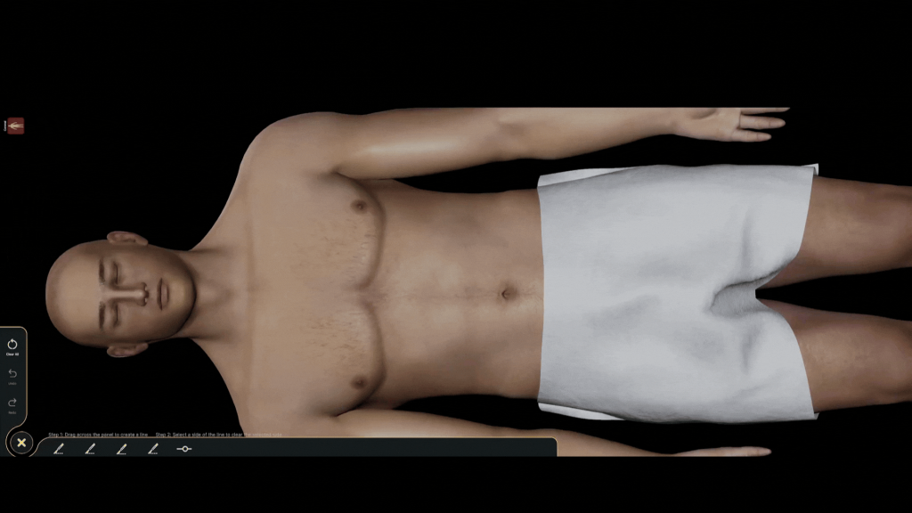
The platform supports sagittal, coronal, and transverse views. Tools such as rotation, pinning, annotation, screenshots, and split-screen viewing help students examine anatomical relationships more closely. DICOM visualisation also allows learners to study anatomical structures alongside corresponding medical imaging.
CADAVIZ extends beyond conventional gross anatomical dissection. It includes regional and organ anatomy, block and window dissection, prosection, embryology, radiology, histology, histopathology, osteology, physiology, muscle movement, and clinical examination.
It also includes presets, quizzes, and OSPE-based assessment. These features help educators structure learning activities, focus attention on particular anatomical regions, assess structure identification, and prepare students for practical examinations.
Because students can revisit regions, manipulate structures, examine spatial relationships, and test their understanding, CADAVIZ can be used before or after cadaveric sessions, during guided teaching, for independent study, and for clinical correlation.
Why Should Medical Colleges Consider Virtual Dissection?
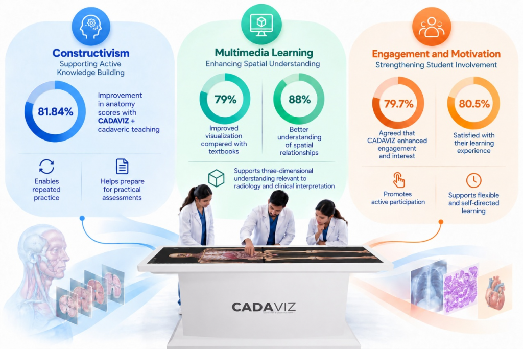
Answer capsule: Virtual dissection strengthens anatomy education by supporting active learning, spatial understanding, engagement, flexible revision, and better laboratory preparation.
The case for virtual dissection table goes beyond adding another digital teaching tool. Its value comes from how well it complements an existing anatomy curriculum, reinforces learning, and gives students more ways to engage with anatomical knowledge across the course.
How Does It Support Active Knowledge Building?
Constructivist learning emphasises that students build knowledge actively rather than passively receive information. Virtual dissection supports this by letting learners explore anatomy, test observations, and revisit challenging regions at their own pace.
In an evaluation of CADAVIZ among undergraduate medical students, the greatest improvement in learning outcomes was reported in the group receiving combined CADAVIZ and cadaveric teaching, with an 81.84% improvement in scores. This supports virtual dissection as a complementary resource within blended anatomy education. Rajan et al. (2025)
Availability outside scheduled teaching also matters. Students can repeat practice, reinforce difficult concepts, and prepare for practical assessments without waiting for the next laboratory session.
Can Virtual Dissection Improve Spatial Understanding?
Yes. Interactive visual learning can help students understand complex anatomical concepts, especially three-dimensional and spatial relationships. The Cognitive Theory of Multimedia Learning provides a useful framework for this type of learning.
In a study of 128 first-year MBBS students, 79% felt CADAVIZ improved anatomical visualisation compared with textbooks, while 88% believed its interactive 3D features improved understanding of spatial relationships. Sreenivasan (2026)
This becomes especially important as students move from foundational anatomy toward radiological interpretation, clinical reasoning, and procedure planning. Seeing a structure in isolation is different from understanding its position, depth, neighbours, and clinical relevance.
Does Virtual Dissection Improve Engagement?
It can. Technology-supported approaches can promote active participation when students are given clear tasks, faculty guidance, and clinically relevant activities.
This aligns with Experiential Learning Theory, where meaningful learning develops through active experience, reflection, and application. In one study, 79.7% of students agreed or strongly agreed that CADAVIZ enhanced their engagement and interest, while 80.5% were satisfied or very satisfied with their experience. More et al. (2025)
Flexible access beyond scheduled classes can also encourage self-directed learning. Students can prepare before practicals, revise after them, and explore areas they found difficult during laboratory teaching.
Virtual anatomy can also strengthen laboratory preparedness. When students familiarise themselves with a region beforehand, scheduled laboratory time can be used more purposefully. Integration with radiology, pathology, surgery, and clinical cases further helps students connect foundational anatomy with later stages of medical education.
How to Integrate the Platform into the Curriculum
Answer capsule: Start with curriculum outcomes, then use virtual dissection for pre-laboratory preparation, guided teaching, post-laboratory revision, assessment, and clinical correlation.
Introducing a virtual dissection platform does not require replacing established anatomy teaching. The more effective approach is to identify where virtual anatomy adds educational value and connect it directly to the learning outcomes of each module.
For colleges considering CADAVIZ or another virtual anatomy platform, implementation should begin with the curriculum and not the technology.
Start With Learning Outcomes, Not Technology
Before introducing virtual dissection, review the expected learning outcomes for every anatomy module. Identify the structures students need to recognise, the anatomical relationships they need to understand, the clinical applications they should interpret, and the competencies that will be assessed.
Virtual activities can then be mapped directly to those outcomes. For example, a neuroanatomy module could include 3D views, cross-sectional anatomy, pathway tracing, and clinical cases.
This keeps the technology in its proper place: a means of achieving defined educational outcomes rather than an objective in itself.
Preparing Students Before They Enter the Anatomy Lab
A virtual dissection platform can prepare students before they enter the cadaver laboratory. A short pre-laboratory activity might include structure identification, anatomical orientation, layer exploration, terminology review, and a preparation quiz.
This gives students early exposure to the region and helps them identify areas that need additional preparation. They can then approach cadaveric teaching with greater familiarity and confidence.
Using CADAVIZ During Lectures and Lab Demonstrations
Virtual anatomy becomes more effective when students receive a defined learning task instead of being asked simply to explore a digital model. CADAVIZ can be incorporated into lectures, tutorials, small-group teaching, laboratory demonstrations, and faculty-led discussions.
Useful activities include:
- Tracing the course of a nerve
- Following a blood vessel through a region
- Comparing superficial and deep structures
- Identifying relationships between organs
- Explaining the anatomical basis of a clinical sign
The distinction matters. “Explore the upper limb” is an instruction to use technology. “Trace the course of the radial nerve and identify its anatomical relationships” is an anatomy learning activity.
Helping Students Revise After Cadaveric Sessions
Learning does not need to end when the anatomy laboratory session ends. Post-laboratory virtual activities help students revisit structures that were difficult to identify or relationships that remained unclear during physical dissection.
Students can re-examine difficult regions, complete targeted activities, answer clinical anatomy questions, and identify topics requiring further revision. Faculty can also use student responses to spot common learning gaps and provide focused reinforcement.
Build Assessment Into Virtual Dissection
Answer capsule: Use virtual quizzes before, during, and after teaching to check preparedness, reinforce anatomical relationships, and identify revision needs.
Assessment should be built into virtual anatomy activities so students can check their understanding while faculty monitor progress against learning outcomes. CADAVIZ includes built-in quizzes that assess anatomical structures and concepts within the platform.
Faculty can also create and assign customised quizzes aligned with specific topics, teaching sessions, curriculum objectives, or practical examination requirements.
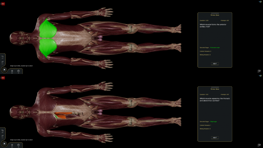
These assessments can be used at different stages:
- Before teaching: Check preparedness before laboratory sessions
- During teaching: Reinforce structure identification and anatomical relationships
- After teaching: Consolidate learning and highlight areas for further study
Custom assessments may include structure-identification questions, anatomical relationship questions, layer-based activities, image- or model-based questions, case-based clinical anatomy questions, and practical examination preparation.
Virtual assessment should complement rather than replace practical assessment. Colleges should continue using cadaveric specimens, physical models, radiological images, demonstrations, and clinical scenarios. The aim is to determine whether students can identify, interpret, explain, and apply anatomical knowledge—not merely recognise a digital model.
Connect Virtual Anatomy With Clinical Cases
One of the most valuable uses of virtual dissection is connecting anatomy with clinical reasoning. A shoulder injury case, for example, could ask students to identify affected structures, trace relevant nerves or vessels, explain functional consequences, and relate anatomical findings to clinical signs.
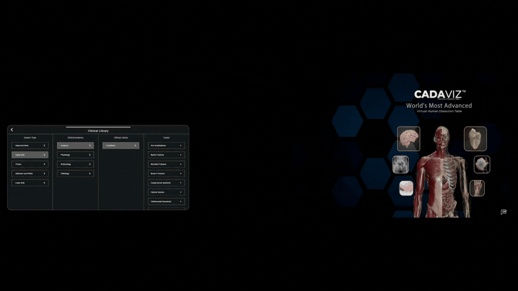
This shifts the question from “Where is the structure?” to “Why does the structure matter clinically?” It helps students connect foundational anatomy with diagnosis, disease, procedures, and treatment.
Use the Platform Across Different Learning Formats
Virtual dissection should not be limited to one teaching session. Its role can change according to the stage of learning.
It can support pre-laboratory preparation, lectures, guided tutorials, small-group activities, independent revision, post-laboratory reinforcement, practical examination preparation, clinical anatomy discussions, and case-based learning.
Early activities may focus on orientation and identification. More advanced activities can emphasise anatomical relationships, interpretation, and clinical application. This makes virtual dissection an adaptable part of the anatomy curriculum rather than a separate digital activity.
Give Students a Clear Learning Roadmap
Access to a virtual dissection platform does not automatically produce effective independent learning. Students should know what they are expected to achieve from each activity.
Each virtual anatomy activity can specify:
- The topic
- Learning outcomes
- Structures to examine
- Recommended completion time
- Questions to answer
- Assessment criteria
This provides academic direction while retaining flexibility for independent 3D exploration. Optional extension activities can let students investigate topics in greater depth.
Prepare Faculty and Students for Virtual Anatomy
Technology is only one part of successful implementation. Faculty and students need to understand how the platform fits into the broader anatomy learning process.
Faculty preparation should cover platform navigation, development of outcome-based activities, integration with teaching sessions, use of assessments, interpretation of student performance, and feedback. Faculty should also participate in curriculum planning and platform evaluation.
Student orientation should cover accessing the platform, navigating anatomical models, using labels and search functions, completing assigned activities, attempting quizzes, reviewing feedback, and obtaining technical support.
Most importantly, students should understand that virtual dissection complements rather than replaces cadaveric dissection, laboratory teaching, lectures, clinical learning, and independent study.
When aligned with learning outcomes, teaching activities, assessment, and clinical application, virtual dissection becomes part of a continuous cycle: preparation, exploration, practice, assessment, feedback, and reinforcement.
A Practical Roadmap for Medical Colleges
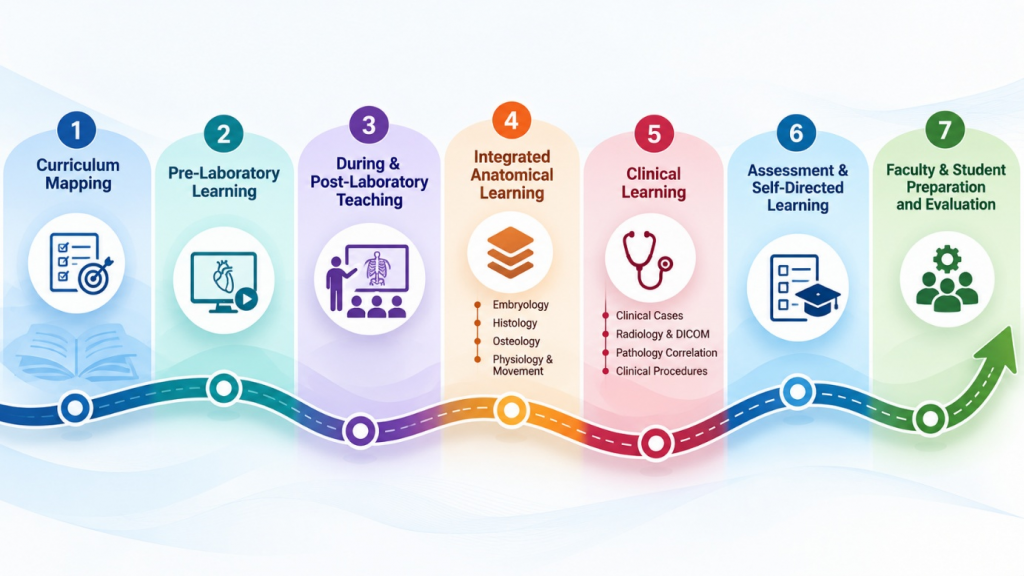
Answer capsule: A phased rollout helps colleges move from curriculum mapping to pre-laboratory preparation, teaching integration, clinical learning, assessment, and evaluation.
A phased approach helps medical colleges integrate virtual dissection while progressively using its different educational capabilities. The aim is not simply to introduce a digital tool, but to establish where virtual anatomy can complement lectures, cadaveric dissection, laboratory teaching, clinical learning, and independent study.
Phase 1: Begin With Curriculum Mapping
Start with the existing anatomy curriculum and its learning outcomes, not the technology. For each module, faculty can identify what students need to know, which structures they should identify, which anatomical relationships they should understand, and which clinical applications they should recognise.
Phase 2: Use Virtual Activities Before Dissection
The platform can introduce a body region before students enter the anatomy laboratory. Faculty can use organ anatomy, regional anatomy, layer exploration, prosection, and anatomical presets to familiarise students with key structures and relationships.
Short structure-identification or preparation quizzes can check foundational knowledge. Students then enter the cadaver laboratory with greater familiarity, allowing virtual learning to function as a pre-laboratory preparation tool.
Phase 3: Use the Platform During and After Laboratory Teaching
During lectures, tutorials, small-group teaching, and laboratory demonstrations, faculty can use virtual dissection to illustrate structures that are difficult to demonstrate through conventional teaching alone.
Students should receive specific exploration tasks, such as tracing a nerve or artery, comparing superficial and deep structures, or locating a structure in relation to surrounding anatomy.
After laboratory sessions, the same region can be revisited virtually. Students can complete short activities, answer clinical questions, and revisit areas requiring further study. This creates continuity between laboratory experience and independent revision.
Phase 4: Progress From Anatomy to Integrated Learning
Once students are comfortable with core virtual dissection, the platform can be used across its broader anatomical and biomedical content. Embryology, histology, histopathology, osteology, physiology, muscle movement, kinesiology, radiology, and DICOM visualisation can be integrated where relevant.
The DICOM viewer offers an additional opportunity to create patient-derived 3D anatomical models. Faculty can demonstrate anatomical variations and clinically relevant configurations that may not be routinely encountered during cadaveric dissection.
Students can compare these models with standard anatomy and see how anatomical variation appears in real patient imaging.
Phase 5: Connect Anatomy With Clinical Learning
The platform can then move students from anatomical identification to clinical application through clinical cases, pathology–radiology correlation, clinical procedures, endoscopic visualisation, cardiovascular physiology, and interactive movement simulations.
For example, a shoulder injury case can require students to identify affected muscles, nerves, vessels, and anatomical spaces, then explain how their relationships contribute to the resulting clinical signs.
This prevents virtual anatomy from becoming simple label memorisation. Instead, it connects structure, function, pathology, imaging, and clinical presentation.
Phase 6: Integrate Assessment and Self-Directed Learning
Assessment should run throughout the learning sequence. Faculty can use built-in assessment resources and create their own quizzes based on the topics and learning outcomes being taught.
These can support pre-laboratory preparation, formative assessment, post-laboratory reinforcement, clinical case discussions, and practical examination revision. Where appropriate, assessment should also include cadaveric specimens, physical models, radiological images, clinical scenarios, and specimen-based examinations.
The platform can also support structured self-directed learning. Faculty can provide clear activity instructions, recommended completion time, questions, and expected outcomes, while optional activities give students additional practice.
Phase 7: Prepare Faculty and Students and Evaluate Implementation
Successful implementation requires orientation for both faculty and students. Faculty should understand not only platform functions but also how to incorporate virtual activities into teaching, create quizzes, use presets, review student performance, and provide feedback.
Students should receive an introductory orientation on navigating models, completing assigned activities, and using the platform for independent learning.
Colleges can evaluate implementation through participation, activity completion, assessment performance, student feedback, faculty experience, time requirements, and technical or accessibility issues. These findings can guide refinement and show where additional virtual activities provide genuine educational value.
Through this phased approach, a virtual dissection platform can evolve from a 3D anatomy resource into a longitudinal learning platform. It can support preparation, exploration, laboratory reinforcement, anatomical variation, imaging, clinical application, self-directed learning, and assessment—while continuing to complement cadaveric dissection and other established anatomy education methods.
Conclusion
Virtual dissection can strengthen anatomy education by extending learning beyond scheduled laboratory sessions. By allowing students to explore structures from multiple angles, it improves spatial understanding and creates opportunities for repeated practice of complex regions at their own pace.
It can also improve preparation for laboratory sessions, helping students become familiar with structures before cadaveric dissection and use laboratory time more efficiently. Flexible access supports revision beyond scheduled teaching, while virtual resources provide additional learning opportunities when cadavers, specimens, or laboratory capacity are limited.
Integration with radiology, surgery, pathology, physical examination, and clinical cases connects anatomical knowledge with clinical practice. Most importantly, virtual dissection should remain aligned with defined learning outcomes so that technology serves a clear educational purpose rather than becoming an isolated demonstration.
When thoughtfully integrated, platforms such as CADAVIZ can complement cadaveric dissection and contribute to a continuous cycle of preparation, exploration, practice, assessment, and clinical application.
Frequently Asked Questions
Can virtual dissection replace cadaveric dissection?
No. Virtual dissection complements cadaveric teaching by supporting preparation, visualisation, revision, clinical integration, and repeated practice. It does not fully replicate tactile learning, manual dissection skills, ethical learning experiences, or the variability of physical specimens.
Is virtual dissection useful for first-year medical students?
Yes. It can support anatomical orientation, body planes, layers, spatial relationships, and basic structure identification. Faculty guidance and curriculum integration remain important for effective use.
How should a medical college choose a virtual dissection platform?
Consider anatomical accuracy, curriculum coverage, usability, accessibility, assessment capabilities, research and publication opportunities, technical support, data privacy, licensing, and total cost. Platforms that support educational research, data collection, outcome evaluation, and publishable studies can also strengthen an institution’s academic ecosystem.
How can virtual dissection be used in anatomy assessments?
It can support formative quizzes, structure identification, guided activities, case-based questions, revision, and practical examination preparation. These should complement, rather than replace, cadaveric, specimen-based, clinical, and radiological assessments.
Does virtual dissection improve student engagement?
It can encourage interaction and repeated practice, but engagement depends on activity design, faculty guidance, curriculum integration, accessibility, usability, and clinical relevance. The technology itself should not be assumed to guarantee improved learning.
When should students use virtual dissection in the anatomy curriculum?
Students can use it before laboratory sessions for preparation, during teaching for guided exploration, and after laboratory sessions for revision. It is also useful for clinical case discussions, imaging correlation, and practical examination preparation.
What is the best way to avoid passive use of virtual anatomy?
Give students specific, outcome-based tasks. For example, ask them to trace a nerve, compare superficial and deep structures, identify anatomical relationships, or explain the anatomical basis of a clinical sign.
How can CADAVIZ support clinical correlation?
CADAVIZ can connect anatomy with clinical cases, radiology, pathology, surgery, physical examination, and DICOM imaging. This helps students move from identifying structures to understanding their clinical relevance.