Dissection is a major focus in the first two years of undergraduate medical education and plays a key role in the area of medicine. It broadens understanding of fundamental medical terminology and touch-and-feel-mediated human body awareness.
Dissection benefits the students in ways that go beyond anatomical learning, results and into their future clinical years. It gives students a realistic perspective of the human body. Future doctors value the cadaver in the dissecting room as their first patient. Over the past decade, we have seen tremendous technological progress permeating every part of our life. Medical education is no exception.
The widespread use of computers, easy access to the internet, and the growing number of software and websites specific to the study of anatomy have revolutionized how students acquire knowledge about it. We will look at the benefits of virtual cadaver dissections in this blog.
Let’s peek into the history of Cadaver Dissection!

Dissection comes from the Latin verb dissecure, which means “to cut into pieces.” Ancient Greek physicians learned considerable information about the human body and health over a long period of time. Dissection of human cadavers has long been an important component of anatomy instruction. The first sanctioned dissection took place at the University of Paris in 1407. Human cadavers have generally been used as a teaching resource since Andreas Vesalius, an anatomist and physician, began dissecting them in 1514.
Why Real Cadaver Dissection?
Students gain a deeper understanding of human anatomical variation and the three-dimensionality of the human body through cadaver dissection. Medical students have a stronger connection to the human body and acquire the empathy needed to become future doctors. The first visual and tactile exposure to the “human body and life” is provided via cadaver dissection to aspiring future doctors.
Real Cadaver Dissection: The Challenges!
The trend of dissection among students has recently changed for various reasons. Beginning medical students, like most laypeople, can hesitate to “cut up” the dead. Additionally, it’s still common for students to faint during the initial dissection in the anatomy labs. Medical students have to bear the foul smell of the cadaver in the dissection hall. Preserving the cadaver is also challenging, and exposure to the preservative chemicals may cause health hazards.
What is the Source of Cadaver?
In the past, anatomists used remains from executions, jails, or poorhouses; but in the 1960s and 1970s, a practical substitute—body donation or the deceased’s informed consent—became available. The sources of human tissue used in medical research and teaching depend on local legislation, program awareness and acceptability, and “unclaimed” bodies, or those of persons who pass away without having close friends or family claim for burial or those who lack financial means to pay for a funeral. When a country is short on corpses, anatomists import cadavers from other countries.
An era of virtual revolution!

The discussions about creating more modern tools for teaching anatomy, such as virtual anatomy, computer-assisted learning, etc., continue to be upbeat. The most effective suggestion for preventing responses from cadaver exposure would be to explain the process beforehand. Additionally, pre-recorded video segments showcasing pertinent images and prior exposure to organs can help medical students cope with the stress in the dissection hall.
With the advent of computer-based learning in the classroom and virtual dissection methods, real cadaver dissection is a thing of the past. With the emergence of web-based technology and the rapid development in the accessibility of educational software and information databases via the internet, computer-aided instruction (CAI) is becoming an important component of modern educational systems.
If, by chance, the students missed any little details during the real dissection, the virtual dissection that goes along with it enables them to remember them.
Students who have watched dissection simulations in advance are better prepared intellectually and psychologically for actual dissection. They gain knowledge of the landmarks, cuts, and structures that need to be highlighted.
Why choose virtual cadaver dissection?
- Accessibility and Flexibility: Virtual cadaver dissection allows medical students to access and study anatomical structures from anywhere at any time. This flexibility is particularly beneficial for distance learning, self-paced study, or accommodating diverse schedules.
- Realistic Representation: Advanced 3D rendering and interactive technologies provide a lifelike representation of human anatomy. Virtual cadavers often mimic the texture, colour, and appearance of real tissues, enhancing the learning experience and providing a high level of realism.
- Enhanced Visualization: Students can manipulate virtual cadavers to view different anatomical structures from multiple angles, zoom in and out, and remove specific layers of tissues. This enhanced visualization helps them better understand complex anatomical relationships.
- Incorporation of Pathological Conditions: Virtual cadaver platforms can simulate various pathological conditions, allowing students to explore and learn about diseased tissues and organs. This fosters a deeper understanding of disease processes and their impact on the human body.
- Risk-Free Learning: Unlike traditional cadaver dissection, virtual dissection eliminates the potential risks associated with handling and exposure to chemicals used for preservation. It also eliminates concerns about ethical considerations related to using human cadavers.
- Repeatability: Students can repeat virtual dissections as many times as needed to reinforce their understanding and improve their anatomical knowledge without the constraints of a physical cadaver.
- Interactive Learning: Virtual dissection often includes interactive quizzes, labelling exercises, and real-time feedback, engaging students in an active learning process that enhances retention and comprehension.
- Incorporation of Multimedia: Virtual platforms can integrate multimedia elements such as videos, animations, and audio explanations, providing additional context and a multi-sensory learning experience.
- Cost-Effective: Setting up and maintaining a physical cadaver lab can be expensive due to the cost of cadavers, preservation chemicals, and storage facilities. Virtual cadaver dissection reduces these costs significantly.
- Global Collaboration: Virtual cadaver dissection facilitates collaboration among medical students and professionals worldwide. It allows individuals from different locations to work together on the same anatomical models and share knowledge and insights.
- Less Time-Consuming: Students can focus specifically on areas they find challenging or need additional practice, saving time compared to the linear nature of traditional dissection.
- Sustainable and Ethical: As concerns about environmental sustainability and ethical considerations grow, virtual cadaver dissection offers a more sustainable and ethically acceptable approach to anatomy education.
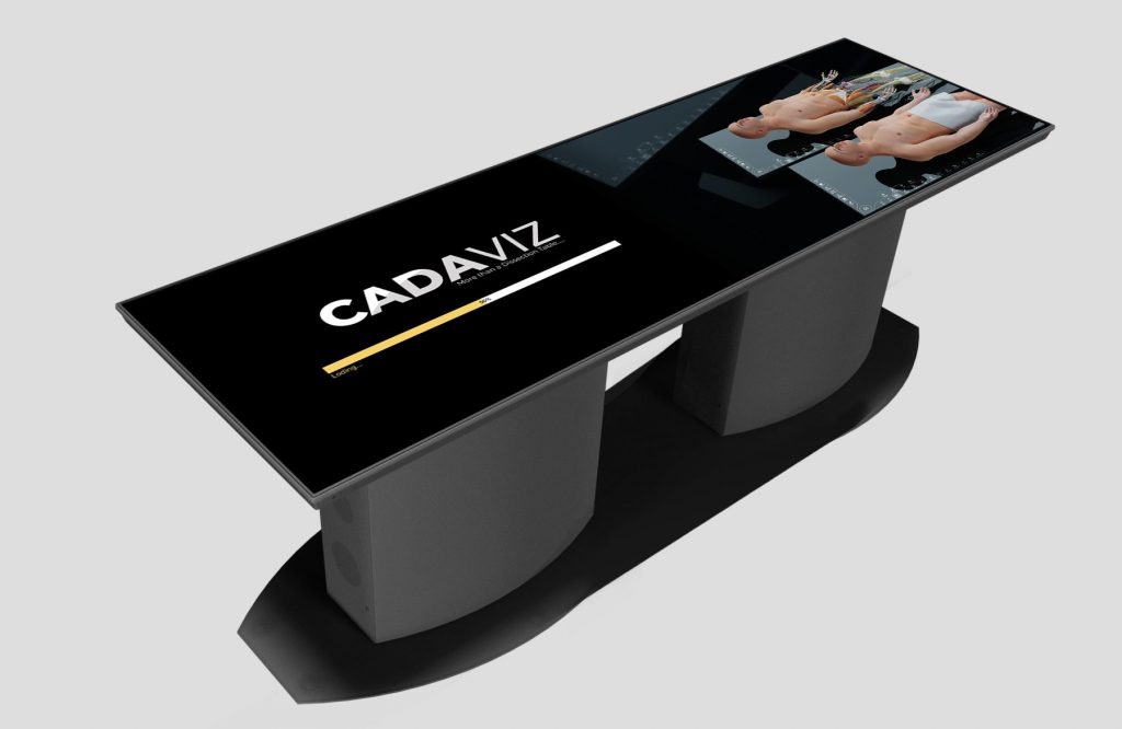
One such virtual cadaver dissection system recommended by medical professionals is CADAVIZ -India’s first and most advanced virtual cadaver dissection table. It enables students to study anatomy in an immersive way, exploring the minute anatomical details of the human body.

Concluding Words!
We may conclude from this reading that cadaver dissection is the foundation for the whole medical curriculum. However, computer-aided virtual cadaver dissection serves as a reinforcement tool, which improves the repeatability of the anatomical information acquired by physical dissection, much like a mega-structure requires several extra pillars and supports for strength and durability.
The greatest way to learn about human anatomical features and for students to do better on tests is to properly combine the two dissection approaches.
CADAVIZ – India’s first and most sophisticated virtual cadaver dissection table is recommended to study virtual cadaver dissection in detail and to excel in medical studies.
Writer – Sneha Adsule
Team Lead Subject Matter Expert-Biology

